Skip to content
Chip Nanoimaging
  • Home
  • TechnologyExpand
    • Chip-Based TIRF
    • Super-Resolution Microscopy
    • 3D Imaging
    • Publications
  • Products
  • ApplicationsExpand
    • Exocytosis
    • Actin Remodeling
    • Tissue Sections
    • Microbiology
    • Gallery
  • News & EventsExpand
    • News
    • Events
  • About usExpand
    • Management
    • Board of directors
    • Investor Relations
    • Contact
Chip Nanoimaging

Gallery

  • [1] EPI fluorescent image of bone marrow cells (U-2 OS) stained with Alexa Fluor 647 Phalloidin to visualize the actin network. Imaging was performed using a 60x NA 1.2 W objective lens. Image 5 is the corresponding TIRF image.
  • [2] TIRF image of the membrane of the commensal E. coli bacteria. The membrane was stained with mCLING-ATTO 647N, and images were acquired with a 60x NA 1.2 W objective lens.
  • [3] TIRF image of the membrane of the commensal E. coli bacteria. The DNA was stained with Hoechst, and images were captured using a 60x NA 1.2 W objective lens.
  • [4] TIRF image of human epithelial cells (A431) stained CellMask Orange, a plasma membrane dye. The cell membrane surface was visualized using a 60x NA 1.2 W objective lens.
  • [5] TIRF image of bone marrow cells (U-2 OS) stained with Alexa Fluor 647 Phalloidin to visualize the actin network. Imaging was performed with a 60x NA 1.2 W objective lens. Image 1 is the corresponding EPI image.
[6] EPI fluorescent image of the actin network in bone marrow cells (U-2 OS) stained with Alexa Fluor 647 Phalloidin. Imaging was performed using a 10x NA 0.1 objective lens.
 TIRF image of the actin network in bone marrow cells (U-2 OS) stained with Alexa Fluor 647 Phalloidin. Imaging was performed using a 10x NA 0.1 objective lens.
  • [7] EPI fluorescent image of the actin network in bone marrow cells (U-2 OS) stained with Alexa Fluor 647 Phalloidin. Imaging was performed using a 10x NA 0.1 objective lens.
[8] TIRF image of a fish intestine tissue sample stained for DNA with Hoechst. The image was acquired using a 25x NA 0.8 W objective lens. Scale bar: 100 µm.
Composite TIRF image of a fish intestine tissue sample stained for actin filaments (magenta) and DNA (green) using Alexa Fluor 647 Phalloidin and Hoechst, respectively. The image was acquired using a 25x NA 0.8 W objective lens. Scale bar: 100 µm.
TIRF image of a fish intestine tissue sample stained for actin filaments with Alexa Fluor 647 Phalloidin. The image was acquired using a 25x NA 0.8 W objective lens. Scale bar: 100 µm.
  • [9] EPI fluorescent image of the actin network in a confluent layer of human epithelial cells (A431) stained with Alexa Fluor 647 Phalloidin. Imaging was performed using a 20x NA 0.45 objective lens.
  • [10] 3D projection (EPI fluorescence) of bone marrow cells (U-2 OS) stained for actin (orange) and DNA (blue). Alexa Fluor 647 Phalloidin and Hoechst were used for staining.
  • [11] Super-resolved reconstruction of the actin network in bone marrow cells (U-2 OS) using fluorescence fluctuation super-resolution microscopy. An image stack of 30 frames was acquired with a 25x NA 0.8 W objective lens. Alexa Fluor 647 Phalloidin was used.
  • [12] Time-lapse using TIRF imaging demonstrating actin remodeling in cells genetically modified to express LifeAct. Images were acquired using a 60x NA 1.2 W objective lens. Scale bar: 40 µm.
  • [13] EPI fluorescent image of bone marrow cells (U-2 OS) stained with Alexa Fluor 647 Phalloidin to visualize the actin network. Imaging was performed using a 60x NA 1.2 W objective lens. The corresponding TIRF image is shown to the right.
  • [14] TIRF image of bone marrow cells (U-2 OS) stained with Alexa Fluor 647 Phalloidin to visualize the actin network. Imaging was performed using a 60x NA 1.2 W objective lens. The corresponding EPI image is shown to the left.
Chip NanoImaging AS
ultra-large field-of-view MICROSCOPY.

Quick Links

  • Home
  • Technology
  • Products
  • Applications
  • News
  • Events
  • About us
Address
CHIP NanoImaging AS
Fiolvegen 15
N-9016 Tromsø
Norway

Enterprise no: NO923780688MVA
Registered in Foretaksregisteret, Norway.

Contact

© 2025 Chip Nanoimaging AS - Read our privacy policy

YouTube Linkedin

Jan Alan Alfheim

Jan A. Alfheim is a business executive with over thirty-five years’ experience bringing product ideas and technology to the chemical and pharmaceutical markets, from product concept inception through discovery and development phases to final marketing campaign and launch. He has direct experience in research, project management, business development & partnering, company start-ups, and product launches.

Alfheim comes from Oncoinvent As where he was Chief Executive Officer from 2016 until 2023. In addition to his role as CEO at Oncoinvent he has led Nordic Nanovector as CEO and has held senior management positions including Chief Business Officer at Clavis Pharma, President of StemPath Inc, Director of Business Development at Neurochem Inc and Project Director at Nycomed Imaging.

Jon Kristian Hagene

With over 35 years of experience in the high-tech photonics industry, Jon Kristian Hagene brings a wealth of expertise to the microscopy field. Originally trained in microelectronics, he has contributed to groundbreaking advancements in optical and spectroscopic gas monitoring, underwater optical imaging, in-line pipeline inspection, and space systems.

Jon’s career includes pivotal roles at Norsk Elektro Optikk, where he served as Research Scientist, Chief Technology Officer (CTO), Acting CEO, and Chief Scientist. From 2019 to 2024, he led Chip NanoImaging AS as CEO, driving innovation and commercialization in cutting-edge imaging solutions.

As a co-founder of NEO Monitors AS, Jon played a key role in its growth and successful acquisition by Nederman Holding for NOK 402 million in 2017. He served on the board of directors of NEO Monitors from its inception in 2003 until the company’s exit.

Jon has also held numerous leadership and advisory positions, including Vice Chairman of the Board of Norsk Elektro Optikk Holding AS (2008–2018) and Board Member of Norsk Elektro Optikk AS (2014–2015, 2016–2018). He has contributed to build equity as a board member in the charitable foundation Irma Salo Jæger og Tycho Jægers Stiftelse (2007–2018).

Øystein I. Helle

Dr. Øystein Ivar Helle is a physicist with over 10 years of experience in integrated optics, photonic technologies, and optical microscopy. As CSO and co-founder of Chip NanoImaging AS, he is recognized for his contributions to photonic chip-based platforms, enabling scalable and high-resolution imaging for applications in biology and pathology.

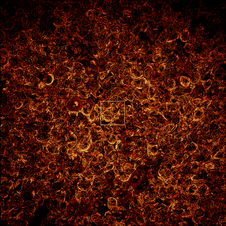
His work addresses key challenges in microscopy, such as limited fields of view and throughput, advancing the boundaries of super-resolution techniques. Dr. Helle has co-authored at least 20 scientific publications and, along with his colleague Professor Balpreet Singh Ahluwalia, was a recipient of the Tycho Jæger Prize in Electro-optics.

His innovative research continues to make a significant impact on life sciences and bioimaging, establishing him as a notable figure in optical nanoscopy.

Balpreet S. Ahluwalia

Professor Balpreet Singh Ahluwalia is a distinguished researcher with about 20 years of R&D experience in nanophotonics, optical microscopy, integrated photonics and bioimaging.

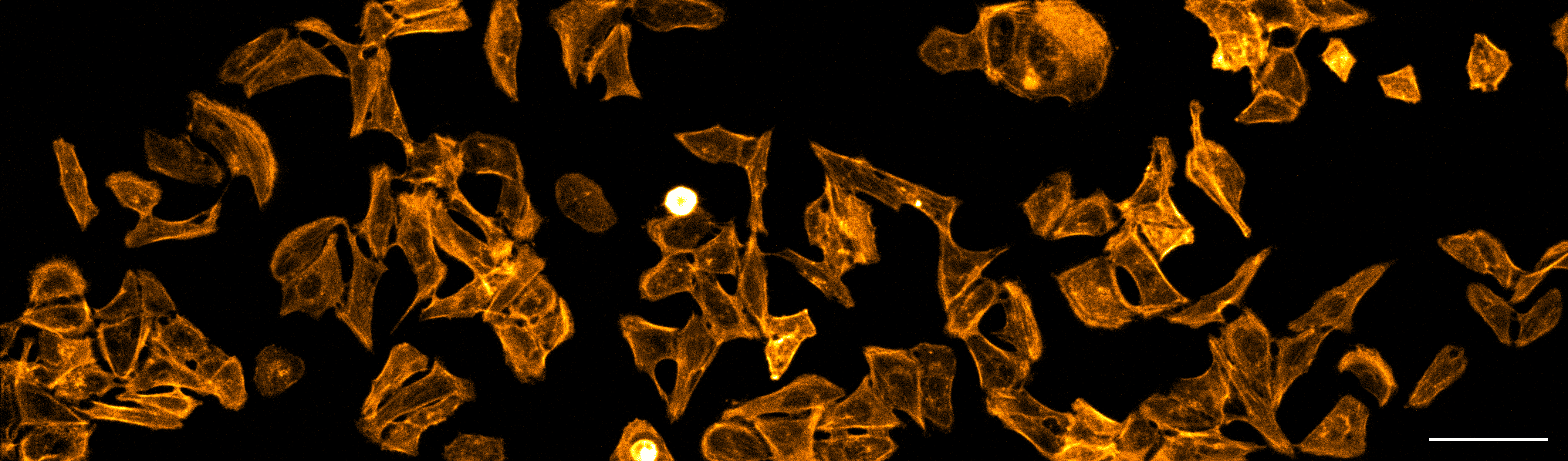
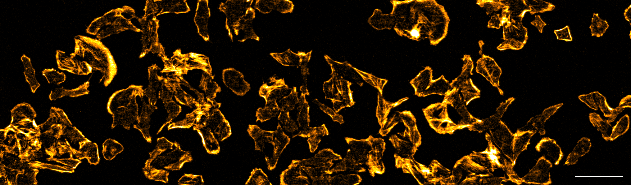
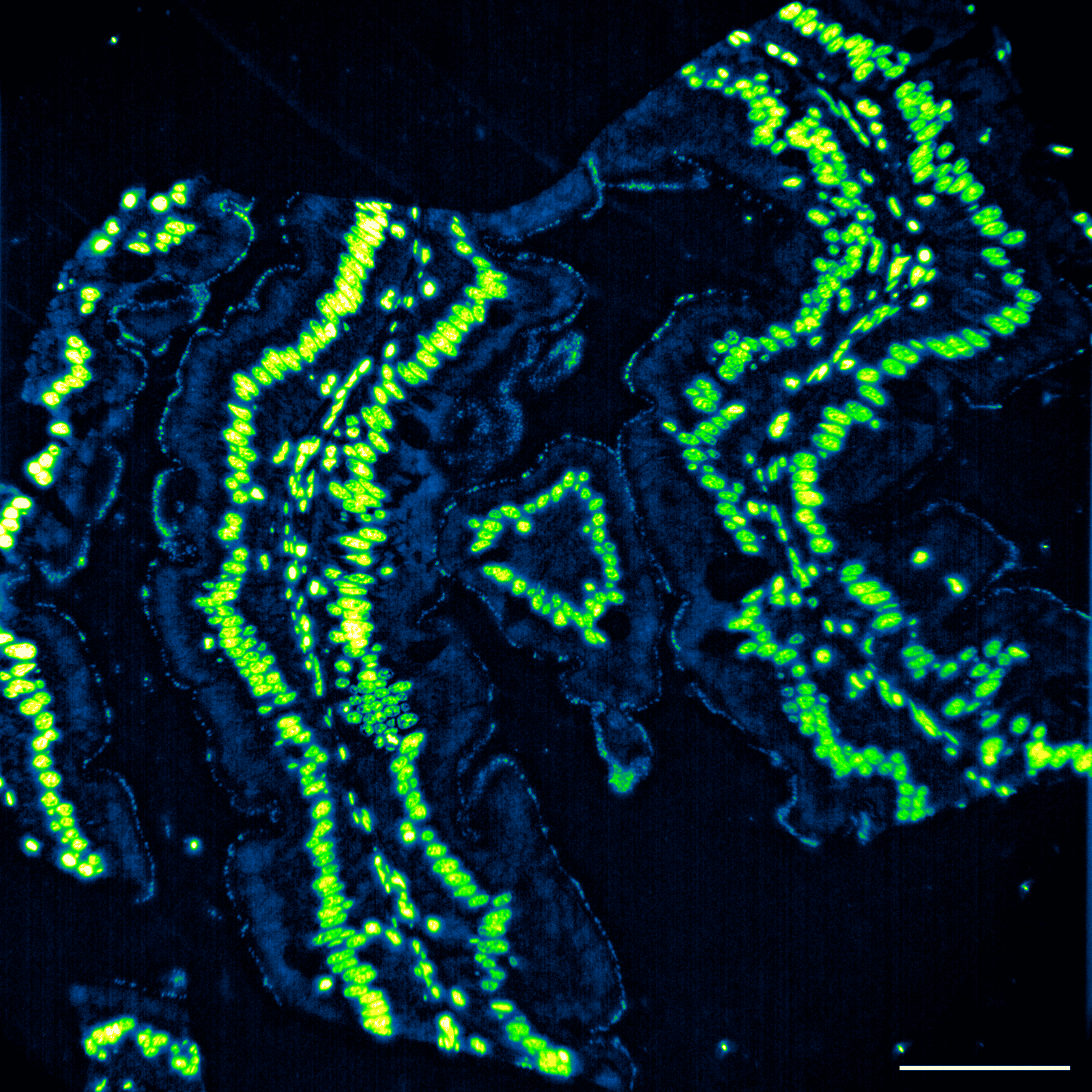
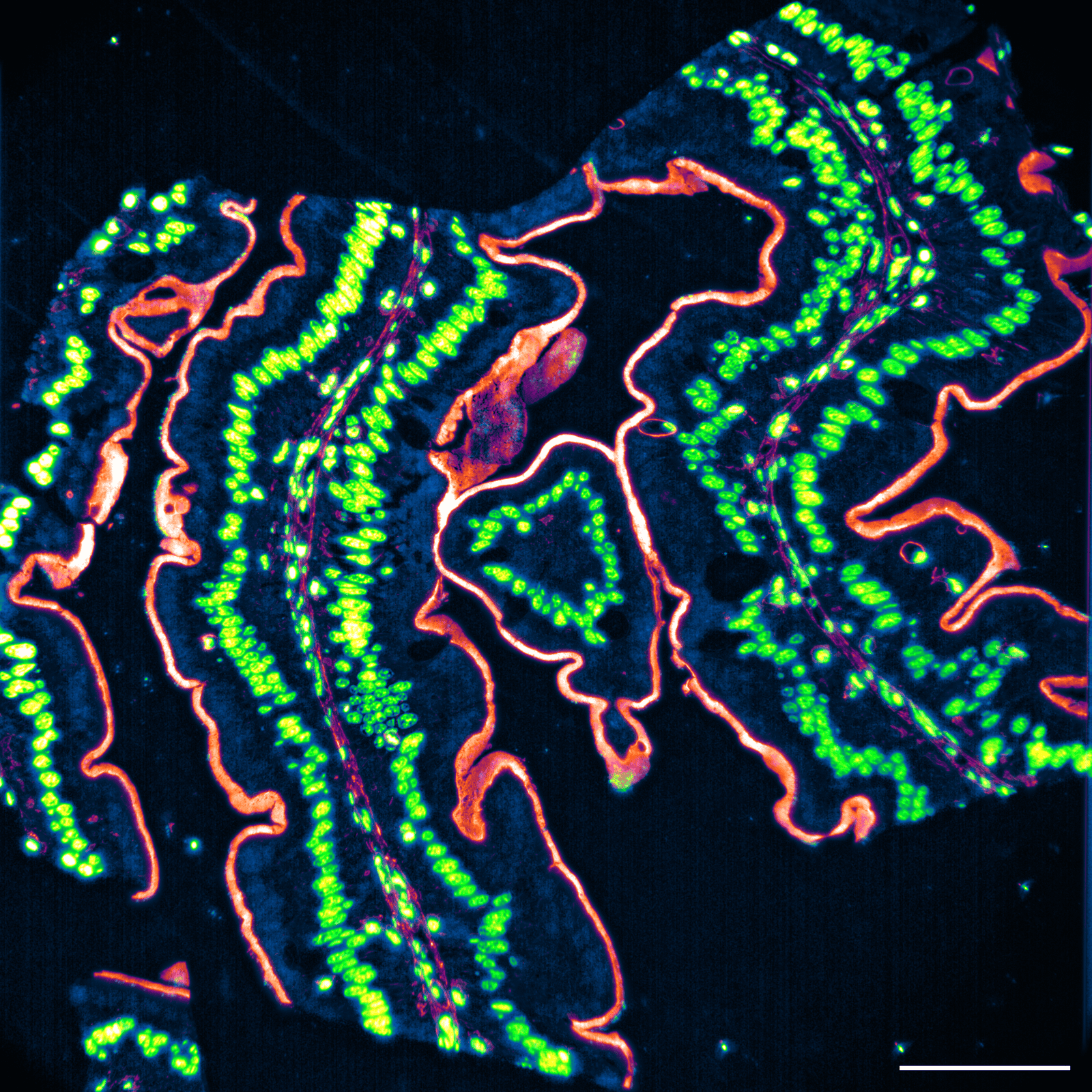
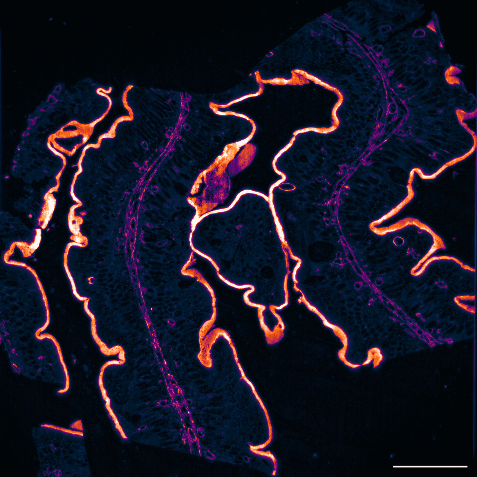
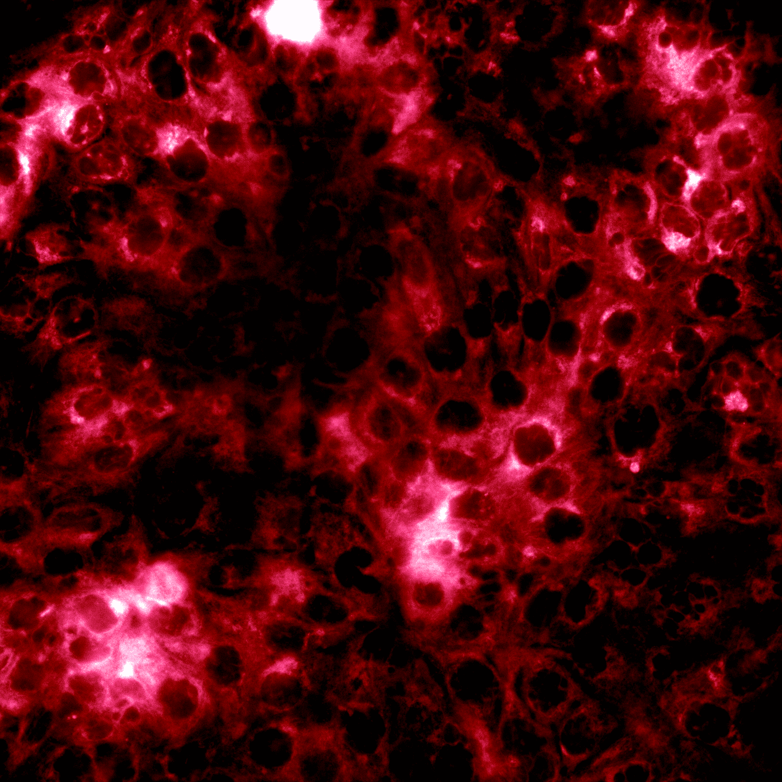
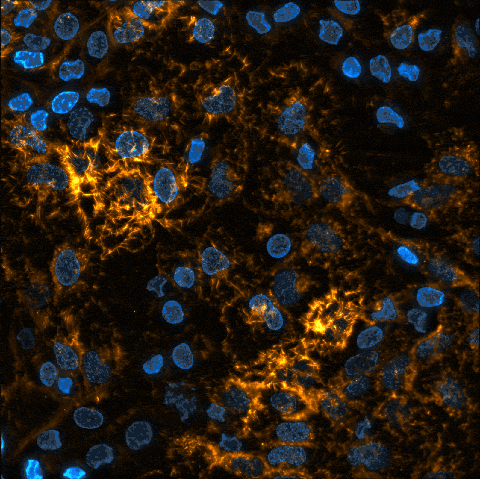
His recent expertise covers the development of advanced super-resolution imaging techniques, enabling high-resolution visualization of biological processes. He is widely recognized for his pioneering work in chip-based super-resolution microscopy, a groundbreaking method that enhances optical resolution while ensuring high-throughput, multi-modality and scalability. He is inventor of on-chip nanoscopy and also co-founder of Chip NanoImaging AS.

Ahluwalia’s innovations have made significant contributions to life sciences and biology, earning him accolades from peers, such as UiT R&D Award, Tycho Jager Prize in Electro-optics (Norway). With an extensive research portfolio spanning across three continents (India, Singapore, USA, UK, and Norway), he has published over 150 scientific research articles and conference papers, cementing his reputation as a leading figure in his field.

Scroll to top
  • Home
  • Technology
    • Chip-Based TIRF
    • Super-Resolution Microscopy
    • 3D Imaging
    • Publications
  • Products
  • Applications
    • Exocytosis
    • Actin Remodeling
    • Tissue Sections
    • Microbiology
    • Gallery
  • News & Events
    • News
    • Events
  • About us
    • Management
    • Board of directors
    • Investor Relations
    • Contact